

Interpreting Complex Genomic Findings in Cardiovascular Disease: Lessons from Three Adult CardioSeq Cases
Cardiovascular disease (CVD) remains the leading cause of death worldwide, affecting hundreds of millions of individuals and accounting for nearly 20 million deaths annually. In the United States, nearly half of adults over the age of 20 are living with some form of CVD. Yet, despite the well-established role of genetic factors to many cardiovascular conditions and professional society recommendations supporting genetic testing, the use of genetic testing in routine cardiovascular care cremains limited.
This gap matters. Large-scale analyses of electronic health records have shown that among 1.7 million CVD patients followed over five years, only ~1.1% underwent any form of genetic testing. As genomic technologies become increasingly capable of detecting clinically meaningful variation, real-world examples are essential to demonstrate both the value and the complexity of genetic testing in cardiovascular medicine. Here, we share insights gained via the CardioSeq Clinical Trial, where patients presenting with cardiovascular disease underwent comprehensive genetic testing that utilized a whole-genome sequencing backbone and focused on a panel of high-confidence genes.
The following three clinical cases highlight how genome-informed testing can uncover clinically relevant findings, while also illustrating the interpretive complexity that arises when multiple variant types, inheritance models, and clinical contexts intersect.
Case 1: When Mendelian Disease and Genetic Risk Coexist
Case 1 illustrates how comprehensive testing can identify more than one clinically meaningful genetic finding in the same individual. In this case, one pathogenic variant was identified in a gene associated with the cardiovascular condition for which the patient was referred for testing, while a second finding was identified as a secondary finding associated with an increased risk of kidney disease.
TTR p.Val142Ile: A well-characterized pathogenic variant
A previously curated missense variant in TTR (p.Val142Ile) was identified and is classified as pathogenic. This variant is a well-established cause of transthyretin amyloidosis and is supported by multiple lines of evidence:
- PS3 (strong), due to functional studies demonstrating amyloidogenicity
- PS4 (strong), increased prevalence in affected individuals, applied with consideration of reduced penetrance and late onset
- Additional supporting evidence from prior reports and gene characteristics
The clinical impact of this finding is important. The variant leads to misfolded transthyretin protein, amyloid fibril formation, and amyloid deposition in multiple tissues. In the heart, this can manifest as ventricular wall thickening, conduction abnormalities, arrhythmias, and heart failure. Importantly, disease is systemic, with possible neurologic, gastrointestinal, renal, and musculoskeletal involvement (Ruberg).
Notably, this variant is relatively common in certain African and Black populations and is not fully penetrant. Carrier status is associated with increased risk of heart failure and mortality, highlighting the continuum between monogenic disease and risk modification, and reinforcing the need for careful interpretation and clear communication of clinical relevance.
While the TTR variant helps explain cardiovascular risk in this patient, it is not the only clinically relevant finding identified. Comprehensive genome sequencing also reveals a second genetic risk factor in APOL1, illustrating how a single test can uncover multiple, independent findings that each require their own clinical and interpretive considerations.
APOL1 Risk Genotypes: Population Genetics Meets Clinical Interpretation
The same individual also carries biallelic risk alleles in APOL1—a compound heterozygous combination of G1 and G2 variants.
Analysis revealed:
- Two missense variants in tight linkage disequilibrium (G1)
- A separate in-frame deletion (G2) on the alternate haplotype
- Distinct haplotypes confirmed by mutually exclusive read patterns
The APOL1 finding adds another layer of complexity. The G1 and G2 alleles are thought to reflect evolutionary selection in certain human populations because they confer resistance to Trypanosoma infection, a parasitic disease, wherein ingestion of APOL1 protein can cause the parasites to lyse. However, these alleles also increase risk for kidney disease, including focal segmental glomerulosclerosis (FSGS) and hypertension-associated end-stage kidney disease (H-ESKD). Importantly, individual alleles do not confer the same disease risk, underscoring why phasing and haplotype interpretation are critical.
Key takeaway: This case demonstrates how a comprehensive approach to genetic testing can uncover multiple clinically relevant findings in a single patient, even when those findings have distinct biological mechanisms and disease implications. Interpreting these findings requires not only variant classification, but also an understanding of penetrance, ancestry, disease mechanism, and how different genetic findings may contribute to overlapping or distinct clinical risks.
Case 2: When Variant Mechanism Challenges Standard Interpretation
Atypical Frameshift in CSRP3
Case 2 demonstrates how genome sequencing can uncover variants that do not fit neatly into standard interpretive categories. The proband carries a frameshift variant in CSRP3, a gene encoding muscle LIM protein, critical for cardiac and skeletal muscle function. The gene is definitively associated with hypertrophic cardiomyopathy (HCM) with a semi-dominant inheritance pattern.
At first glance, the variant appears to be a frameshift. However, unlike a typical frameshift that leads to truncation, this variant rewrites much of the coding sequence and results in C-terminal extension. Because the variant disrupts both LIM domains, which are essential for protein function, classification requires careful consideration of the underlying disease mechanism rather than reliance on variant type alone. Although mechanistically atypical, variant classification incorporates:
- PVS1 (strong) due to disruption of critical functional domains
- PM2 (supporting) for rarity
- PM5 (supporting) an unconventional application
PM5 is generally reserved for missense variants where a different pathogenic variant has been observed at the same residue. However, in this case, multiple (n=11) pathogenic/likely pathogenic frameshift variants have been reported in the same region of CSRP3, which also resulted in LIM domain disruption and the same protein extension. Based on this established disease mechanism and precedent in variant databases, PM5 is applied at reduced strength. This combination of evidence (one strong, two supporting) supports a Likely Pathogenic classification Strengthen Case 1 transitions so the reader understands how the TTR and APOL1 findings relate (or don’t relate) clinically.
This type of adaptation of PM5 is not without precedent. In other gene-disease contexts, laboratories have extended PM5 beyond its original definition when supported by strong biological rationale. For example, in BRCA1 and BRCA2, PM5 has been applied (with varying strengths) to nonsense variants occurring within well-characterized functional exons, reflecting the consistent pathogenic loss of function mechanism of those regions. While these applications remain context-dependent and are not universally standardized, they illustrate how gene-specific knowledge can inform thoughtful modification of ACMG/AMP criteria.
This approach reflects a broader reality in genomic interpretation: guidelines provide an essential framework, but variant interpretation often requires thoughtful, gene-specific adaptation when the mechanism is biologically plausible and supported by prior evidence.
Borderline STR Expansion in DMPK
The same individual also harbors a CAG repeat expansion in the 3′ UTR of DMPK, the gene associated with myotonic dystrophy type 1.
- Genome sequencing data: 55 repeats
- Orthogonal testing: 49 repeats (±2 repeat uncertainty)
This repeat-length result places the variant right at the boundary between the premutation range and the fully penetrant pathogenic range. Clinical interpretation is further complicated by the correlation between repeat length, age of onset, and disease severity, where patients with a lower number of repeats (yet still in the pathogenic range) may have late-onset, milder disease that typically includes arrhythmia
Key takeaway: Some variants require interpretation beyond standard categories. Atypical frameshifts, borderline repeat expansions, and other complex findings demand integration of technical evidence, gene-specific biology, and clinical context.
Case 3: Mosaic Structural Variation and the Challenge of Secondary Findings
Case 3 involves a 4.6 Mb mosaic deletion on chromosome 13, encompassing the RB1 gene, a well-known tumor suppressor.
Key observations:
- Reduced sequencing coverage across the region
- Absence of uniform hemizygosity, suggesting mosaicism rather than a germline deletion
- Clean breakpoints supporting a true structural event
While RB1 loss is classically associated with early-onset retinoblastoma, mosaic deletions in this region present a unique interpretive challenge. Although similar alterations have been observed in hematologic malignancies, mosaic chromosomal changes can also occur as age-related clonal hematopoiesis in otherwise healthy individuals Given its inclusion on the ACMG secondary findings list, the variant affecting RB1 was reported, but with atypical zygosity and it has uncertain clinical significance in this context.
Key takeaway: Mosaic structural variants require careful consideration of tissue origin, age-related changes, and clinical context. When identified incidentally, especially in genes associated with secondary findings, interpretation must balance the potential clinical relevance with uncertainty about tissue origin and disease association.
Broader Lessons for Cardiovascular Genomics
Together, these cases illustrate how the task of variant interpretation has evolved in the genome sequencing era:
- Expanding Variant Classes, Expanding Complexity. Genome sequencing enables the detection of more variant types. However, these variants, e.g. repeat expansions and mosaic CNVs, can introduce unique challenges to variant interpretation.
- Beyond Binary Classification. Not all variants fit into simple frameworks. Risk alleles, reduced penetrance variants, borderline expansions, and mosaic findings may require nuanced interpretation.
- Importance of Visualization and Manual Review. Read-level inspection remains critical for confirming variant quality, determining phase (e.g., APOL1), and identifying mosaicism.
- Flexibility in Applying Guidelines. The ACMG/AMP framework provides an essential structure for rigorous variant classification, but context-dependent adaptation may be necessary, particularly for non-canonical variant effects.
Conclusion
Genome sequencing is a powerful tool for uncovering the genetic basis of disease. Because guideline-recommended genetic testing is underutilized in cardiovascular disease, a large number of genetic etiologies likely remain undiagnosed. In particular, tests leveraging the power of whole genome sequencing can uncover a wider range of variant types and thereby provide answers—and often personalized treatment recommendations—to more patients. However, with this power comes increased interpretive complexity. As these cases demonstrate, variant interpretation increasingly requires integration of diverse data types, careful application of guidelines, and the ability to decipher complexity. Nevertheless, variant interpretation scientists are keeping pace with advances in sequencing technology to deliver clinically meaningful insights to patients.
References
-
Palaniappan, L. P., Allen, N. B., Almarzooq, Z. I., Anderson, C. A., Arora, P., Avery, C. L., … & American Heart Association Council on Epidemiology and Prevention Statistics Committee and Stroke Statistics Committee. (2026). 2026 Heart Disease and Stroke Statistics: A Report of US and Global Data From the American Heart Association. Circulation.
-
Musunuru, K., Hershberger, R. E., Day, S. M., Klinedinst, N. J., Landstrom, A. P., Parikh, V. N., … & American Heart Association Council on Genomic and Precision Medicine; Council on Arteriosclerosis, Thrombosis and Vascular Biology; Council on Cardiovascular and Stroke Nursing; and Council on Clinical Cardiology. (2020). Genetic testing for inherited cardiovascular diseases: a scientific statement from the American Heart Association. Circulation: Genomic and Precision Medicine, 13(4), e000067.
-
Wilde, A. A., Semsarian, C., Márquez, M. F., Shamloo, A. S., Ackerman, M. J., Ashley, E. A., … & Deneke, T. (2022). European heart rhythm association (EHRA)/Heart rhythm society (HRS)/Asia pacific heart rhythm society (APHRS)/Latin American heart rhythm society (LAHRS) expert consensus statement on the state of genetic testing for cardiac diseases. Europace, 24(8), 1307-1367.
-
Longoni M, Bhasin K, Ward A, Lee D, Nisson M, Bhatt S, Rodriguez F, Dash R. Real-world utilization of guideline-directed genetic testing in inherited cardiovascular diseases. Front Cardiovasc Med. 2023 Oct 17;10:1272433. doi: 10.3389/fcvm.2023.1272433. PMID: 37915745; PMCID: PMC10616303.
-
Bui, Q. M., Morales, A., Adler, E. D., Urey, M. A., Sinha, A., Wang, J., … & Ramsis, M. (2026). Real World Genetic Testing Practices in Cardiomyopathy: National insights from 1.7 million patients. Journal of Cardiac Failure.
-
Amendola, L. M., Coffey, A. J., Lowry, J., Avecilla, J., Malhotra, A., Chawla, A., … & Taft, R. J. (2025). Development of a comprehensive cardiovascular disease genetic risk assessment test. Genetics in Medicine Open, 103482.
-
Ruberg, F. L., & Maurer, M. S. (2024). Cardiac amyloidosis due to transthyretin protein: a review. Jama, 331(9), 778-791.
-
Parcha, V., Malla, G., Irvin, M. R., Armstrong, N. D., Judd, S. E., Lange, L. A., … & Arora, P. (2022). Association of transthyretin Val122Ile variant with incident heart failure among black individuals. Jama, 327(14), 1368-1378.
-
Friedman, D. J., & Pollak, M. R. (2020). APOL1 and kidney disease: from genetics to biology. Annual review of physiology, 82(1), 323-342.
-
Vafiadaki, E., Arvanitis, D. A., & Sanoudou, D. (2015). Muscle LIM Protein: Master regulator of cardiac and skeletal muscle functions. Gene, 566(1), 1-7.
-
Germain, P., Delalande, A., & Pichon, C. (2022). Role of muscle LIM protein in mechanotransduction process. International Journal of Molecular Sciences, 23(17), 9785.
-
Parsons, M. T., de la Hoya, M., Richardson, M. E., Tudini, E., Anderson, M., Berkofsky-Fessler, W., … & Spurdle, A. B. (2024). Evidence-based recommendations for gene-specific ACMG/AMP variant classification from the ClinGen ENIGMA BRCA1 and BRCA2 Variant Curation Expert Panel. The American Journal of Human Genetics, 111(9), 2044-2058.
-
Bird TD. Myotonic Dystrophy Type 1. 1999 Sep 17 [updated 2024 Nov 14]. In: Adam MP, Bick S, Mirzaa GM, Pagon RA, Wallace SE, Amemiya A, editors. GeneReviews® [Internet]. Seattle (WA): University of Washington, Seattle; 1993–2026. PMID: 20301344.
-
Machiela, M. J., Zhou, W., Caporaso, N., Dean, M., Gapstur, S. M., Goldin, L., … & Chanock, S. J. (2016). Mosaic 13q14 deletions in peripheral leukocytes of non-hematologic cancer cases and healthy controls. Journal of human genetics, 61(5), 411-418.
-
Miller, D. T., Lee, K., Abul-Husn, N. S., Amendola, L. M., Brothers, K., Chung, W. K., … & ACMG Secondary Findings Working Group. (2023). ACMG SF v3. 2 list for reporting of secondary findings in clinical exome and genome sequencing: a policy statement of the American College of Medical Genetics and Genomics (ACMG). Genetics in Medicine, 25(8), 100866.
When a “loss” isn’t a loss: what a de novo GLUL start‑loss variant teaches us about variant interpretation and exome reanalysis
Despite increased knowledge, more than half of detected variants are classified as variants of uncertain significance (VUS), and in turn, more than half of patients undergoing exome or genome sequencing still receive a negative report. Thus, there is tremendous need to improve the interpretation of tricky variants. Start‑loss variants are often assumed to result in protein deletion and are frequently deprioritized during variant interpretation. Emerging evidence now challenges this assumption. A recently characterized de novo start‑loss variant in GLUL demonstrates how disrupted translational regulation can have surprising effects on protein function that drive autosomal dominant disease, and why unresolved exomes deserve systematic reanalysis as gene-disease knowledge evolves.
Presented during Illumina’s Grand Rounds webinar series, this case highlights two key lessons for variant scientists and laboratory geneticists:
- Start‑loss variants are not uniformly loss‑of‑function and may exert pathogenic effects through other mechanisms.
- Reanalysis is often the only path to diagnosis as interpretation frameworks incorporate new biological discoveries.
Why this case matters for variant interpretation
A dominant disease mechanism established only recently
Start‑loss variants are commonly interpreted as null. However, functional studies now show that certain GLUL start‑loss variants give rise to a truncated yet enzymatically active glutamine synthetase that escapes normal glutamine‑dependent degradation. This leads to pathologic glutamine accumulation and downstream neurotoxicity, an effect more consistent with toxic gain‑of‑function via protein stabilization than classic haploinsufficiency (Figure 1).
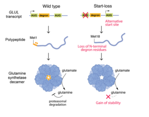
Figure 1 – Functional Consequences of a GLUL Start-Loss Variant
Until 2024, GLUL was not recognized as a cause of autosomal dominant neurodevelopmental disease. The first large series linking heterozygous de novo GLUL variants to Developmental and Epileptic Encephalopathy (DEE) was published only shortly before this case was reanalyzed, fundamentally altering how variants in this gene are triaged and interpreted.1
Clinical summary
Daniel is a five‑year‑old child with severe developmental delay, drug‑resistant epilepsy, and progressive white matter abnormalities. His neurological course includes early hypotonia, developmental regression, multiple seizure types (including myoclonus, absence, and focal seizures with apnea), and profound motor impairment. Brain MRI demonstrated diffuse cerebral atrophy and hypomyelination, and EEG showed disorganized background activity with focal epileptiform discharges.
The constellation of findings suggested a genetic etiology, initially raising suspicion for a metabolic or mitochondrial disorder.
Initial testing and the absence of a diagnosis
Standard genetic testing, including karyotype, chromosomal microarray, and clinical exome sequencing, was performed early in Daniel’s diagnostic journey and returned negative. For four years, no molecular explanation was identified.
Exome reanalysis reveals three variants
In 2024, Daniel’s parents requested reanalysis of his original exome data. Three heterozygous variants were identified:
- GLUL: start‑loss variant (pathogenic, de novo)
- HECW2: missense variant (variant of uncertain significance, inherited)
- HMGCL: stop‑gained variant (pathogenic, carrier state)
Variant interpretation: separating signal from noise
GLUL: the disease‑causing variant
The most compelling finding was a de novo start‑loss variant in GLUL, encoding glutamine synthetase, a critical enzyme in CNS ammonia detoxification and neurotransmitter cycling.
Pathogenicity was supported by multiple orthogonal lines of evidence:
- A disease mechanism now known to be protein‑stabilizing and dominant
- De novo occurrence confirmed by parental testing
- Functional data demonstrating altered regulation rather than loss of enzymatic activity
- Absence of the variant from population databases
- Strong phenotypic concordance with recently reported GLUL‑associated DEE cases
Downstream ATG codons can sometimes rescue translation in start‑loss variants, including the GLUL variants associated with DEE. However, functional data indicated this mechanism produced a truncated GLUL protein with pathological effects. The variant was therefore classified as pathogenic.
GLUL is an emerging autosomal dominant DEE gene whose disease mechanism was only recently established. Critically, this variant would have had an uncertain interpretation and may have never been prioritized before the GLUL disease mechanism was established.
HECW2: plausible but likely not causal
A heterozygous missense variant in HECW2, a gene associated with autosomal dominant neurodevelopmental disorders, initially appeared plausible based on phenotypic overlap. However, inheritance from an unaffected parent, lack of conservation at the altered residue, and absence from ClinVar reduced support for pathogenicity. The variant was classified as a VUS and not considered causative.
HMGCL: pathogenic but not explanatory
A pathogenic stop‑gained variant in HMGCL was also identified. However, HMGCL‑related disease is autosomal recessive and presents with severe neonatal metabolic decompensation. Daniel’s heterozygous status and clinical course were inconsistent with a causal role, and he was classified as an unaffected carrier.
Why the diagnosis was missed initially
Daniel’s case underscores a fundamental reality in clinical genomics: a negative exome reflects the limits of current knowledge, not the absence of a genetic cause.
The diagnostic breakthrough became possible only after peer‑reviewed publications in 2024–2025 established heterozygous GLUL variants as a cause of autosomal dominant DEE and clarified their molecular mechanism. These studies described multiple patients with highly concordant phenotypes and demonstrated transcriptional and regulatory disruption via RNA sequencing. 1,2,3
Broader implications for genomic practice
This case illustrates several field‑wide lessons relevant to variant interpretation and laboratory workflows:
- Start‑loss variants require mechanism‑aware interpretation, not automatic null assumptions.
- Newly established gene-disease relationships can retrospectively unlock diagnoses.
- Systematic exome reanalysis is essential in unresolved neurodevelopmental disorders.
- Family advocacy remains a powerful driver of diagnostic closure.
Because our knowledge of gene-disease relationships continues to expand, genomic data are not static, but rather, are a renewable diagnostic resource. As illustrated by Daniel’s case, reanalysis can transform uncertainty into clarity for families and advance the interpretive practice of the field.
References
-
Jones AG, Aquilino M, Tinker RJ, et al. Clustered de novo start‑loss variants in GLUL result in a developmental and epileptic encephalopathy via stabilization of glutamine synthetase. Am J Hum Genet. 2024;111(4):729‑741.
-
Carbonell E, Stenton SL, Ganesh VS, et al. Male proband with intractable seizures and a de novo start‑codon‑disrupting variant in GLUL. Hum Genet Genomics Adv. 2025;6(2).
-
Oh DE, Jang SS, Kim WJ, et al. Expanding the clinical and genetic spectrum of GLUL‑related developmental and epileptic encephalopathy. Sci Rep. 2025;15(1):35655.